To determine safety, efficacy and mechanism of action of novel therapeutic agents, a comprehensive assessment of pathologic changes is essential for preclinical studies.
EPO´s state-of-the-art histology laboratories are equipped to accommodate a variety of analyses including necropsies and tissue processing, preparation of sections from frozen or paraffin-embedded samples, tissue-based tests and diagnostic services and immunohistochemistry (IHC). Our expert scientists and technicians have developed and validated methodologies to facilitate a broad spectrum of studies and are ready to support you at every stage of the histology workflow.
Necropsy and tissue processing
Necropsy generally includes a complete post-mortem gross examination of organs as well as a microscopic examination (histopathology) of selected tissues, if required. EPO´s necropsy services include the following:
- Supervision of necropsy procedure by a board-certified pathologist
- Tissue collection including whole-body and organ perfusion
- Cryo- or paraffin-embedding of tissues for immunohistochemistry (IHC)
- Tissue collection for molecular diagnostics such as PCR
Histopathology
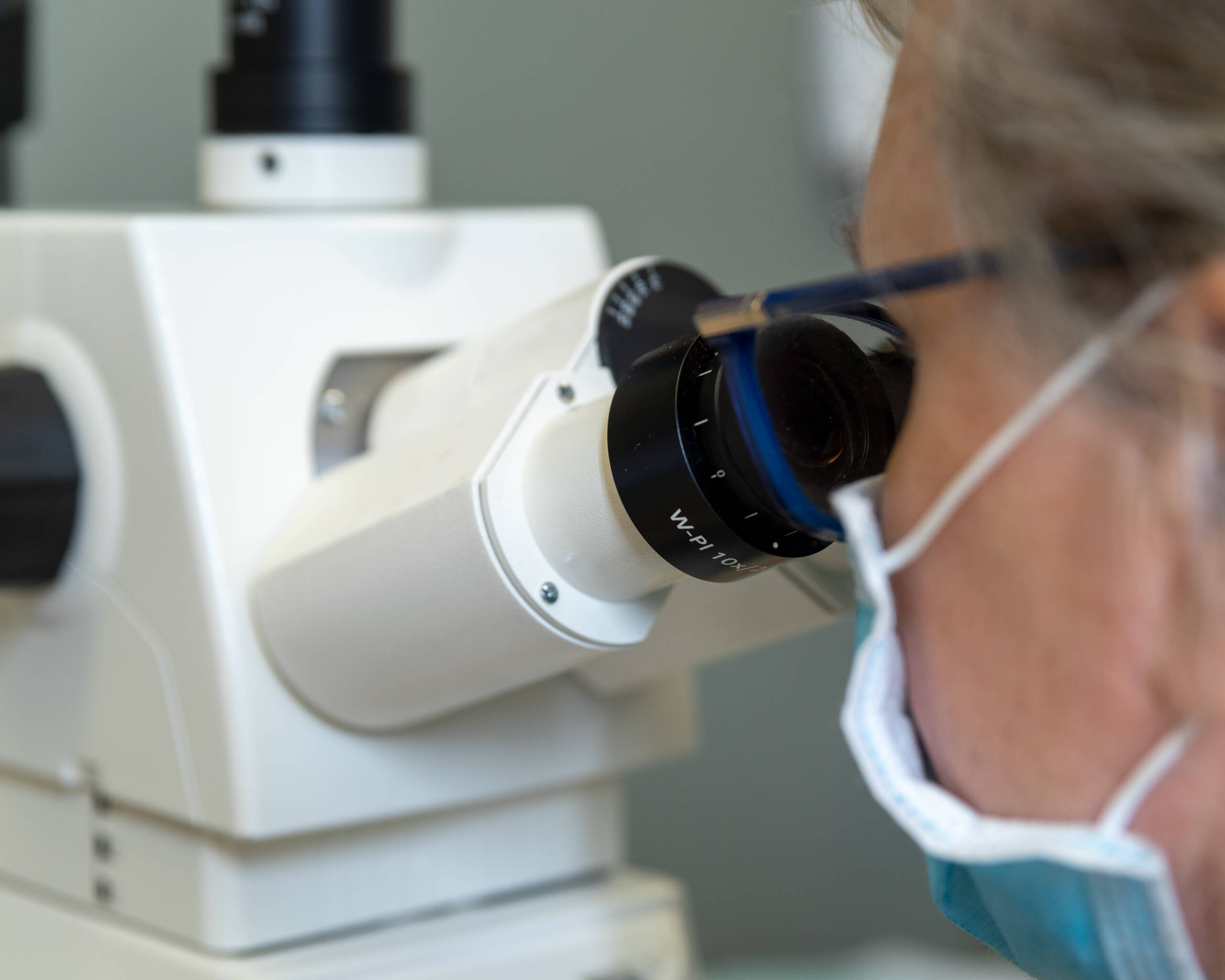
Histopathology (or histology) involves the examination of sampled whole tissues under the microscope. We offer the standard hematoxylin and eosin (H&E) histology including evaluation by a board-certified pathologist. Apart from this, we perform a wide variety of special stains which employ a dye or chemical that has an affinity for the particular tissue component that is to be visualized.
Immunohistochemistry
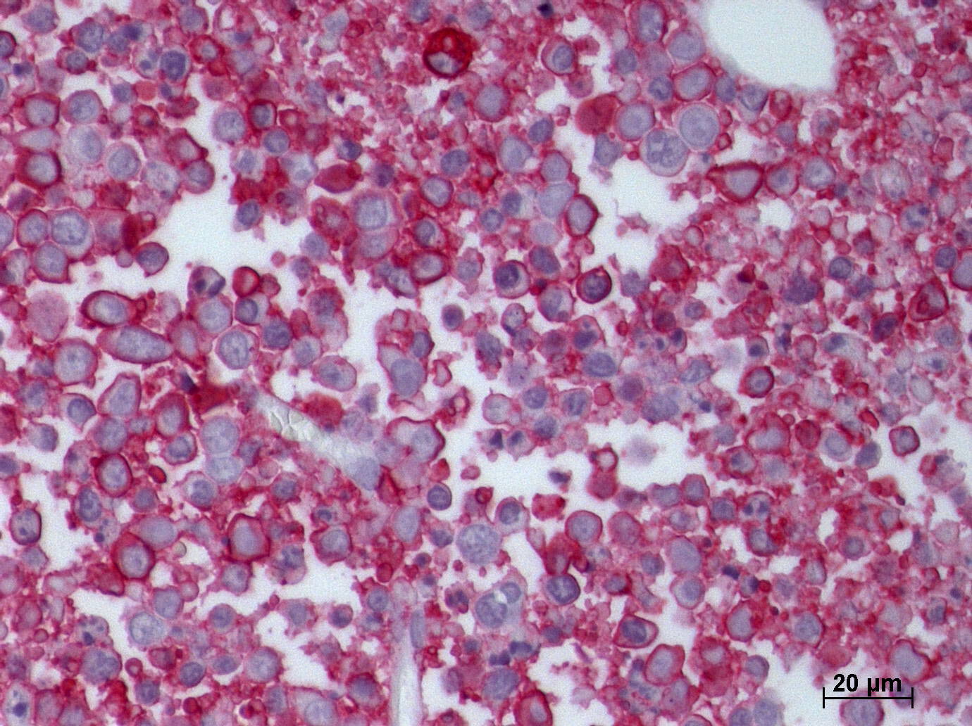
Our expertise also covers state-of-the-art immunohistochemistry (IHC) on both frozen and paraffin sections. IHC utilizes highly specific antibodies for the visualization of target expression patterns and thereby provides quantitative, qualitative and spatio-temporal information on processes that take place in tissues. For this, we already can rely on a constantly growing portfolio of established protocols for the detection of a broad variety of targets. Please contact our pathologist Prof. Dr. Sterner-Kock PhD, DECVP for a complete list of established protocols.
In addition to our standard protocols we are also offering customized IHC services. These services include:
- Antibody selection for your specific study
- Antibody validation with suitable controls
- Staining protocol development and optimization

